Image of the Day
The images that appear in the "Image of the Day" are selected for the freshness of their views on Brain Mapping, their esthetic appeal, their quirkiness, or someimes just to prod you into thinking about the field and its context. Their appearance here is not an endorsement of their subject matter.
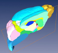 |
Sagittal cut through the 3D microMRI C57BL/6J mouse brain atlas
The image shows in great detail a sagittal cut through the 3D microMRI C57BL/6J mouse brain atlas. Brain structures are labeled in different colors. The structures visible here are neocortex (cyan), olfactory bulb (golden), cerebellum (pink), brain stem (blue), caudate putamen (light green), hippocampus (deep blue), thalamus (milky white), external capsule (yellow), internal capsule (red), globus pallidus (deep pink), amygdala (light yellow), inferior coliculli (yellowish green), superior coliculli (white) and hypothalamus (deep cyan). Image courtesy of Dr. Helene Benveniste and Dr. Yu Ma, Brookhaven National Laboratory and Stony Brook University. Grant NIH R01 EB 00233-04 and P41 RR16105. |
For more information on this image, visit:
[ This Link ]
Submitted by:
Mark Cohen
|