scanSTAT can read images in a variety of common native formats including General Electric (*.MR), Advanced NMR (*.img), MGH formats (*.bshort, *.buchar, *.bfloat) and Mayo Clinic Analyze (*.img) and manages byte ordering where needed.
Tutorial/Demonstration - Viewing Images from a variety of formats
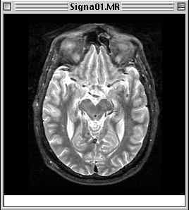
The image you select will open immediately. If you were to open Signa01.MR, for example, you should get a window which looks something like this:
View Image display of Signa01.MR
Note that the gray levels, which are calculated by the program, are initialized well, even for surface coil images (e.g., small.bshort).
You may use the Display menu selection View Options... to alter the brightness and interpolation modes of the image. You may also use the Size To... selection to enlarge or minify the image.
If you did not choose small.bshort, close the current image either by clicking in its close box or selecting File -> Close. Now use File -> View Image... to open small.bshort.
Note that small.bshort is not a single image, but is in fact a fMRI time series of 75 images. When opened using View Image..., however, the statistical analysis features normally used when dealing with time series are unavailable. To use these features, you must inform scanSTAT that you will be working with a time series, by using the Open Time Data... menu item. This is described in the next section of the walkthrough.